Hickened Mass of Cells on the Blastocyst From Which the Baby Will Develop Is Called

The initial stages of human embryonic development (embryogenesis)
Human embryonic development, or homo embryogenesis, is the development and formation of the human being embryo. Information technology is characterised by the processes of cell partitioning and cellular differentiation of the embryo that occurs during the early stages of development. In biological terms, the development of the human body entails growth from a 1-celled zygote to an developed man. Fertilisation occurs when the sperm cell successfully enters and fuses with an egg cell (ovum). The genetic material of the sperm and egg then combine to form a single cell called a zygote and the germinal phase of development commences.[1] Embryonic development in the man, covers the first eight weeks of development; at the showtime of the 9th calendar week the embryo is termed a fetus. Human embryology is the study of this development during the offset eight weeks after fertilisation. The normal period of gestation (pregnancy) is about nine months or 40 weeks.
The germinal stage refers to the time from fertilization through the evolution of the early on embryo until implantation is completed in the uterus. The germinal stage takes around 10 days.[2] During this stage, the zygote begins to carve up, in a process called cleavage. A blastocyst is so formed and implanted in the uterus. Embryogenesis continues with the next stage of gastrulation, when the three germ layers of the embryo grade in a process chosen histogenesis, and the processes of neurulation and organogenesis follow.
In comparison to the embryo, the fetus has more than recognizable external features and a more complete gear up of developing organs. The entire procedure of embryogenesis involves coordinated spatial and temporal changes in factor expression, cell growth and cellular differentiation. A about identical process occurs in other species, specially among chordates.
Germinal phase [edit]
Fertilization [edit]
Fertilization takes place when the spermatozoon has successfully entered the ovum and the 2 sets of genetic textile carried past the gametes fuse together, resulting in the zygote (a single diploid cell). This usually takes place in the ampulla of one of the fallopian tubes. The zygote contains the combined genetic material carried by both the male and female gametes which consists of the 23 chromosomes from the nucleus of the ovum and the 23 chromosomes from the nucleus of the sperm. The 46 chromosomes undergo changes prior to the mitotic segmentation which leads to the formation of the embryo having two cells.
Successful fertilization is enabled by three processes, which also act every bit controls to ensure species-specificity. The first is that of chemotaxis which directs the motion of the sperm towards the ovum.[3] Secondly, an adhesive compatibility between the sperm and the egg occurs. With the sperm adhered to the ovum, the 3rd process of acrosomal reaction takes place; the front end part of the spermatozoan head is capped by an acrosome which contains digestive enzymes to interruption down the zona pellucida and allow its entry.[4] The entry of the sperm causes calcium to be released which blocks entry to other sperm cells.[4] A parallel reaction takes place in the ovum chosen the zona reaction. This sees the release of cortical granules that release enzymes which digest sperm receptor proteins, thus preventing polyspermy.[five] The granules also fuse with the plasma membrane and alter the zona pellucida in such a way as to forbid farther sperm entry.
Cleavage [edit]

The beginning of the cleavage procedure is marked when the zygote divides through mitosis into 2 cells. This mitosis continues and the first two cells divide into iv cells, then into eight cells then on. Each division takes from 12 to 24 hours. The zygote is large compared to whatsoever other cell and undergoes cleavage without any overall increase in size. This means that with each successive subdivision, the ratio of nuclear to cytoplasmic material increases.[6]
Initially, the dividing cells, called blastomeres ( blastos Greek for sprout), are undifferentiated and aggregated into a sphere enclosed within the membrane of glycoproteins (termed the zona pellucida) of the ovum. When eight blastomeres have formed, they begin to develop gap junctions, enabling them to develop in an integrated fashion and co-ordinate their response to physiological signals and environmental cues.[7]
When the cells number around 16, the solid sphere of cells within the zona pellucida is referred to as a morula.[8] At this stage the cells start to bind firmly together in a process chosen compaction, and cleavage continues as cellular differentiation.
Blastulation [edit]
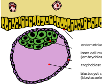
Cleavage itself is the get-go stage in blastulation, the process of forming the blastocyst. Cells differentiate into an outer layer of cells (collectively called the trophoblast) and an inner cell mass. With further compaction the individual outer blastomeres, the trophoblasts, become indistinguishable. They are still enclosed within the zona pellucida. This compaction serves to brand the structure watertight, containing the fluid that the cells will later secrete. The inner mass of cells differentiate to become embryoblasts and polarise at one cease. They shut together and form gap junctions, which facilitate cellular advice. This polarisation leaves a crenel, the blastocoel, creating a structure that is now termed the blastocyst. (In animals other than mammals, this is called the blastula.) The trophoblasts secrete fluid into the blastocoel. The resulting increment in size of the blastocyst causes it to hatch through the zona pellucida, which so disintegrates.[6] This process is called hatching of the homo embryo and it takes identify on the sixth day of embryo development, immediately before the implantation process. The hatching of human embryo is supported by proteases secreted past the cells of the blastocyst, which digest proteins of the zona pellucida, giving ascension to a hole. And so, thanks to the rhythmic expansion and contractions of the blastocyst, an increase of the pressure inside the blastocyst itself occur, the hole expands and finally the blastocyst can sally from this rigid envelope.
The inner cell mass will give ascension to the pre-embryo,[9] the amnion, yolk sac and allantois, while the fetal part of the placenta will form from the outer trophoblast layer. The embryo plus its membranes is called the conceptus, and by this stage the conceptus has reached the uterus. The zona pellucida ultimately disappears completely, and the now exposed cells of the trophoblast let the blastocyst to adhere itself to the endometrium, where it will implant. The formation of the hypoblast and epiblast, which are the two main layers of the bilaminar germ disc, occurs at the beginning of the second week.[10] Both the embryoblast or the trophoblast will turn into two sub-layers.[11] The inner cells will plow into the hypoblast layer, which will surround the other layer, chosen the epiblast, and these layers will class the embryonic disc that will develop into the embryo.[10] [11] The trophoblast will besides develop two sub-layers: the cytotrophoblast, which is in front end of the syncytiotrophoblast, which in turn lies within the endometrium.[10] Next, another layer called the exocoelomic membrane or Heuser's membrane will appear and surroundings the cytotrophoblast, also equally the archaic yolk sac.[11] The syncytiotrophoblast volition abound and volition enter a stage called lacunar stage, in which some vacuoles volition appear and be filled by blood in the following days.[10] [11] The development of the yolk sac starts with the hypoblastic flat cells that course the exocoelomic membrane, which volition coat the inner role of the cytotrophoblast to class the primitive yolk sac. An erosion of the endothelial lining of the maternal capillaries by the syncytiotrophoblastic cells results into formation of the maternal sinusoids from where the claret will begin to penetrate and menstruum into and through the trophoblastic lacunae to give rise to the uteroplacental circulation.[12] [13] Subsequently, new cells derived from yolk sac will be established between trophoblast and exocelomic membrane and will give ascent to extra-embryonic mesoderm, which will form the chorionic cavity.[11]
At the terminate of the second week of development, some cells of the trophoblast penetrate and grade rounded columns into the syncytiotrophoblast. These columns are known as master villi. At the aforementioned time, other migrating cells grade into the exocelomic cavity a new cavity named the secondary or definitive yolk sac, smaller than the archaic yolk sac.[xi] [12]
Implantation [edit]
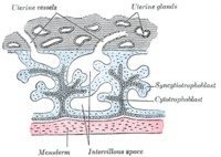
Trophoblast differentiation
After ovulation, the endometrial lining becomes transformed into a secretory lining in preparation of accepting the embryo. Information technology becomes thickened, with its secretory glands becoming elongated, and is increasingly vascular. This lining of the uterine cavity (or womb) is at present known as the decidua, and information technology produces a great number of large decidual cells in its increased interglandular tissue. The blastomeres in the blastocyst are bundled into an outer layer called the trophoblast. The trophoblast so differentiates into an inner layer, the cytotrophoblast, and an outer layer, the syncytiotrophoblast. The cytotrophoblast contains cuboidal epithelial cells and is the source of dividing cells, and the syncytiotrophoblast is a syncytial layer without prison cell boundaries.
The syncytiotrophoblast implants the blastocyst in the decidual epithelium past projections of chorionic villi, forming the embryonic part of the placenta. The placenta develops once the blastocyst is implanted, connecting the embryo to the uterine wall. The decidua here is termed the decidua basalis; it lies between the blastocyst and the myometrium and forms the maternal part of the placenta. The implantation is assisted by hydrolytic enzymes that erode the epithelium. The syncytiotrophoblast also produces human chorionic gonadotropin, a hormone that stimulates the release of progesterone from the corpus luteum. Progesterone enriches the uterus with a thick lining of blood vessels and capillaries so that information technology can oxygenate and sustain the developing embryo. The uterus liberates sugar from stored glycogen from its cells to nourish the embryo.[14] The villi begin to co-operative and contain blood vessels of the embryo. Other villi, called final or complimentary villi, commutation nutrients. The embryo is joined to the trophoblastic shell by a narrow connecting stem that develops into the umbilical cord to attach the placenta to the embryo.[eleven] [xv] Arteries in the decidua are remodelled to increase the maternal blood menstruation into the intervillous spaces of the placenta, assuasive gas commutation and the transfer of nutrients to the embryo. Waste matter products from the embryo will lengthened across the placenta.
Every bit the syncytiotrophoblast starts to penetrate the uterine wall, the inner cell mass (embryoblast) also develops. The inner prison cell mass is the source of embryonic stem cells, which are pluripotent and tin can develop into any i of the three germ layer cells, and which have the dominance to give rise to all the tissues and organs.
Embryonic disc [edit]
The embryoblast forms an embryonic disc, which is a bilaminar disc of ii layers, an upper layer called the epiblast (archaic ectoderm) and a lower layer called the hypoblast (primitive endoderm). The disc is stretched between what volition become the amniotic cavity and the yolk sac. The epiblast is adjacent to the trophoblast and made of columnar cells; the hypoblast is closest to the blastocyst crenel and fabricated of cuboidal cells. The epiblast migrates abroad from the trophoblast downwardly, forming the amniotic cavity, the lining of which is formed from amnioblasts developed from the epiblast. The hypoblast is pushed down and forms the yolk sac (exocoelomic crenel) lining. Some hypoblast cells drift forth the inner cytotrophoblast lining of the blastocoel, secreting an extracellular matrix forth the mode. These hypoblast cells and extracellular matrix are called Heuser's membrane (or the exocoelomic membrane), and they cover the blastocoel to form the yolk sac (or exocoelomic crenel). Cells of the hypoblast drift along the outer edges of this reticulum and form the extraembryonic mesoderm; this disrupts the extraembryonic reticulum. Presently pockets form in the reticulum, which ultimately coalesce to form the chorionic crenel (extraembryonic coelom).
Gastrulation [edit]


Embryo fastened to placenta in amniotic crenel
The primitive streak, a linear ring of cells formed by the migrating epiblast, appears, and this marks the kickoff of gastrulation, which takes identify around the seventeenth 24-hour interval (week three) afterwards fertilisation. The process of gastrulation reorganises the 2-layer embryo into a three-layer embryo, and also gives the embryo its specific caput-to-tail, and forepart-to-back orientation, by way of the archaic streak which establishes bilateral symmetry. A primitive node (or primitive knot) forms in front of the primitive streak which is the organiser of neurulation. A primitive pit forms equally a depression in the centre of the archaic node which connects to the notochord which lies directly underneath. The node has arisen from epiblasts of the amniotic cavity floor, and information technology is this node that induces the formation of the neural plate which serves as the basis for the nervous system. The neural plate will grade opposite the primitive streak from ectodermal tissue which thickens and flattens into the neural plate. The epiblast in that region moves down into the streak at the location of the archaic pit where the process chosen ingression, which leads to the formation of the mesoderm takes place. This ingression sees the cells from the epiblast move into the primitive streak in an epithelial-mesenchymal transition; epithelial cells go mesenchymal stem cells, multipotent stromal cells that tin can differentiate into various jail cell types. The hypoblast is pushed out of the way and goes on to form the amnion. The epiblast keeps moving and forms a 2d layer, the mesoderm. The epiblast has now differentiated into the 3 germ layers of the embryo, and so that the bilaminar disc is now a trilaminar disc, the gastrula.
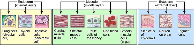
The three germ layers are the ectoderm, mesoderm and endoderm, and are formed as three overlapping flat discs. It is from these iii layers that all the structures and organs of the torso will exist derived through the processes of somitogenesis, histogenesis and organogenesis.[16] The embryonic endoderm is formed by invagination of epiblastic cells that drift to the hypoblast, while the mesoderm is formed past the cells that develop between the epiblast and endoderm. In general, all germ layers will derive from the epiblast.[11] [15] The upper layer of ectoderm will give rising to the outermost layer of skin, key and peripheral nervous systems, optics, inner ear, and many connective tissues.[17] The middle layer of mesoderm volition give rising to the heart and the beginning of the circulatory system equally well every bit the bones, muscles and kidneys. The inner layer of endoderm will serve as the starting point for the evolution of the lungs, intestine, thyroid, pancreas and bladder.
Following ingression, a blastopore develops where the cells have ingressed, in one side of the embryo and it deepens to become the archenteron, the first determinative stage of the gut. As in all deuterostomes, the blastopore becomes the anus whilst the gut tunnels through the embryo to the other side where the opening becomes the rima oris. With a functioning digestive tube, gastrulation is now completed and the side by side phase of neurulation can begin.
Neurulation [edit]

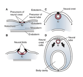
Following gastrulation, the ectoderm gives ascension to epithelial and neural tissue, and the gastrula is now referred to as the neurula. The neural plate that has formed as a thickened plate from the ectoderm, continues to broaden and its ends start to fold upwardly equally neural folds. Neurulation refers to this folding procedure whereby the neural plate is transformed into the neural tube, and this takes place during the fourth week. They fold, along a shallow neural groove which has formed every bit a dividing median line in the neural plate. This deepens as the folds go along to gain summit, when they will meet and shut together at the neural crest. The cells that migrate through the most cranial part of the archaic line form the paraxial mesoderm, which will give rise to the somitomeres that in the procedure of somitogenesis will differentiate into somites that will form the sclerotomes, the syndetomes,[18] the myotomes and the dermatomes to grade cartilage and bone, tendons, dermis (skin), and musculus. The intermediate mesoderm gives rise to the urogenital tract and consists of cells that migrate from the centre region of the archaic line. Other cells migrate through the caudal part of the primitive line and course the lateral mesoderm, and those cells migrating by the most caudal part contribute to the extraembryonic mesoderm.[11] [15]
The embryonic disc begins flat and round, just eventually elongates to take a wider cephalic part and narrow-shaped caudal stop.[10] At the first, the primitive line extends in cephalic direction and 18 days after fertilization returns caudally until it disappears. In the cephalic portion, the germ layer shows specific differentiation at the kickoff of the fourth week, while in the caudal portion it occurs at the terminate of the quaternary week.[xi] Cranial and caudal neuropores become progressively smaller until they shut completely (by twenty-four hour period 26) forming the neural tube.[19]
Development of organs and organ systems [edit]

Organogenesis is the evolution of the organs that begins during the 3rd to eighth week, and continues until nascency. Sometimes total development, as in the lungs, continues after nascency. Dissimilar organs take part in the development of the many organ systems of the body.
Blood [edit]
Haematopoietic stem cells that give rise to all the blood cells develop from the mesoderm. The development of blood formation takes place in clusters of blood cells, known as claret islands, in the yolk sac. Blood islands develop exterior the embryo, on the umbilical vesicle, allantois, connecting stalk, and chorion, from mesodermal hemangioblasts.
In the centre of a blood isle, hemangioblasts form the haematopoietic stem cells that are the forerunner to all types of claret cell. In the periphery of a blood isle the hemangioblasts differentiate into angioblasts the precursors to the blood vessels.[twenty]
Heart and circulatory system [edit]
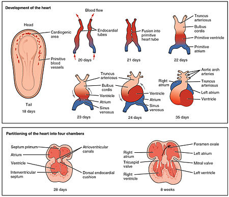
The heart is the start functional organ to develop and starts to beat and pump blood at around 22 days.[21] Cardiac myoblasts and claret islands in the splanchnopleuric mesenchyme on each side of the neural plate, requite rise to the cardiogenic region.[11] : 165 This is a horseshoe-shaped area most to the head of the embryo. Past day 19, following cell signalling, two strands begin to form as tubes in this region, equally a lumen develops inside them. These two endocardial tubes grow and by day 21 have migrated towards each other and fused to form a single archaic eye tube, the tubular heart. This is enabled by the folding of the embryo which pushes the tubes into the thoracic cavity.[22]
Also at the same time that the endocardial tubes are forming, vasculogenesis (the development of the circulatory system) has begun. This starts on day 18 with cells in the splanchnopleuric mesoderm differentiating into angioblasts that develop into flattened endothelial cells. These join to grade small-scale vesicles chosen angiocysts which join up to grade long vessels chosen angioblastic cords. These cords develop into a pervasive network of plexuses in the formation of the vascular network. This network grows by the boosted budding and sprouting of new vessels in the process of angiogenesis.[22] Following vasculogenesis and the evolution of an early vasculature, a stage of vascular remodelling takes place.
The tubular centre quickly forms v distinct regions. From head to tail, these are the infundibulum, bulbus cordis, primitive ventricle, primitive atrium, and the sinus venosus. Initially, all venous blood flows into the sinus venosus, and is propelled from tail to caput to the truncus arteriosus. This will split to form the aorta and pulmonary artery; the bulbus cordis will develop into the right (primitive) ventricle; the primitive ventricle will form the left ventricle; the primitive atrium will become the front end parts of the left and right atria and their appendages, and the sinus venosus will develop into the posterior part of the right atrium, the sinoatrial node and the coronary sinus.[21]
Cardiac looping begins to shape the eye every bit 1 of the processes of morphogenesis, and this completes by the end of the quaternary week. Programmed cell decease (apoptosis) at the joining surfaces enables fusion to have place.[22] In the heart of the fourth week, the sinus venosus receives blood from the three major veins: the vitelline, the umbilical and the common cardinal veins.
During the commencement 2 months of evolution, the interatrial septum begins to form. This septum divides the archaic atrium into a right and a left atrium. Firstly it starts as a crescent-shaped piece of tissue which grows downwards as the septum primum. The crescent shape prevents the consummate closure of the atria allowing claret to be shunted from the right to the left atrium through the opening known as the ostium primum. This closes with farther development of the system but before it does, a 2d opening (the ostium secundum) begins to form in the upper atrium enabling the continued shunting of blood.[22]
A 2d septum (the septum secundum) begins to form to the right of the septum primum. This also leaves a pocket-size opening, the foramen ovale which is continuous with the previous opening of the ostium secundum. The septum primum is reduced to a small flap that acts as the valve of the foramen ovale and this remains until its closure at birth. Between the ventricles the septum inferius also forms which develops into the muscular interventricular septum.[22]
Digestive system [edit]
The digestive system starts to develop from the third calendar week and by the twelfth week, the organs accept correctly positioned themselves.
Respiratory organisation [edit]
The respiratory system develops from the lung bud, which appears in the ventral wall of the foregut almost four weeks into development. The lung bud forms the trachea and two lateral growths known as the bronchial buds, which enlarge at the beginning of the fifth calendar week to course the left and right main bronchi. These bronchi in plough grade secondary (lobar) bronchi; three on the right and 2 on the left (reflecting the number of lung lobes). Tertiary bronchi grade from secondary bronchi.
While the internal lining of the larynx originates from the lung bud, its cartilages and muscles originate from the 4th and 6th pharyngeal arches.[23]
Urinary system [edit]
Kidneys [edit]
Iii dissimilar kidney systems course in the developing embryo: the pronephros, the mesonephros and the metanephros. Simply the metanephros develops into the permanent kidney. All three are derived from the intermediate mesoderm.
Pronephros [edit]
The pronephros derives from the intermediate mesoderm in the cervical region. It is not functional and degenerates earlier the end of the 4th week.
Mesonephros [edit]
The mesonephros derives from intermediate mesoderm in the upper thoracic to upper lumbar segments. Excretory tubules are formed and enter the mesonephric duct, which ends in the cloaca. The mesonephric duct atrophies in females, just participate in evolution of the reproductive arrangement in males.
Metanephros [edit]
The metanephros appears in the 5th week of evolution. An outgrowth of the mesonephric duct, the ureteric bud, penetrates metanephric tissue to form the primitive renal pelvis, renal calyces and renal pyramids. The ureter is besides formed.
Bladder and urethra [edit]
Between the fourth and seventh weeks of development, the urorectal septum divides the cloaca into the urogenital sinus and the anal canal. The upper part of the urogenital sinus forms the bladder, while the lower role forms the urethra.[23]
Reproductive system [edit]
Integumentary organization [edit]
The superficial layer of the skin, the epidermis, is derived from the ectoderm. The deeper layer, the dermis, is derived from mesenchyme.
The formation of the epidermis begins in the second month of development and information technology acquires its definitive organization at the end of the 4th month. The ectoderm divides to form a flat layer of cells on the surface known as the periderm. Further division forms the private layers of the epidermis.
The mesenchyme that will course the dermis is derived from three sources:
- The mesenchyme that forms the dermis in the limbs and body wall derives from the lateral plate mesoderm
- The mesenchyme that forms the dermis in the back derives from paraxial mesoderm
- The mesenchyme that forms the dermis in the face and neck derives from neural crest cells[23]
Nervous system [edit]
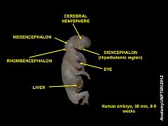
Development of brain in 8-calendar week-old embryo
Late in the quaternary calendar week, the superior office of the neural tube bends ventrally as the cephalic flexure at the level of the future midbrain—the mesencephalon. Above the mesencephalon is the prosencephalon (future forebrain) and below information technology is the rhombencephalon (future hindbrain).
Cranial neural crest cells migrate to the pharyngeal arches as neural stem cells, where they develop in the process of neurogenesis into neurons.
The optical vesicle (which eventually becomes the optic nerve, retina and iris) forms at the basal plate of the prosencephalon. The alar plate of the prosencephalon expands to grade the cognitive hemispheres (the telencephalon) whilst its basal plate becomes the diencephalon. Finally, the optic vesicle grows to course an optic outgrowth.
Development of physical features [edit]
Face and neck [edit]
| | This section needs expansion. You tin assist past calculation to it. (November 2017) |
From the tertiary to the eighth week the face and neck develop.
Ears [edit]
The inner ear, middle ear and outer ear have distinct embryological origins.
Inner ear [edit]
At about 22 days into development, the ectoderm on each side of the rhombencephalon thickens to form otic placodes. These placodes invaginate to form otic pits, and then otic vesicles. The otic vesicles then form ventral and dorsal components.
The ventral component forms the saccule and the cochlear duct. In the sixth week of development the cochlear duct emerges and penetrates the surrounding mesenchyme, travelling in a spiral shape until it forms 2.5 turns by the end of the eighth week. The saccule is the remaining part of the ventral component. Information technology remains connected to the cochlear duct via the narrow ductus reuniens.
The dorsal component forms the utricle and semicircular canals.
Center ear [edit]
The tympanic cavity and eustachian tube are derived from the first pharyngeal pouch (a cavity lined by endoderm). The distal part of the cleft, the tubotympanic recess, widens to create the tympanic cavity. The proximal part of the scissure remains narrow and creates the eustachian tube.
The bones of the middle ear, the ossicles, derive from the cartilages of the pharyngeal arches. The malleus and incus derive from the cartilage of the first pharyngeal arch, whereas the stapes derives from the cartilage of the second pharyngeal arch.
Outer ear [edit]
The external auditory meatus develops from the dorsal portion of the first pharyngeal scissure. Half dozen auricular hillocks, which are mesenchymal proliferations at the dorsal aspects of the first and 2nd pharyngeal arches, form the auricle of the ear.[23]
Eyes [edit]
The eyes begin to develop from the tertiary week to the tenth week.

Movements of embryo at 9 weeks gestational age.
Limbs [edit]
| | This section needs expansion. You can help past adding to information technology. (November 2017) |
At the end of the fourth week limb development begins. Limb buds announced on the ventrolateral aspect of the trunk. They consist of an outer layer of ectoderm and an inner role consisting of mesenchyme which is derived from the parietal layer of lateral plate mesoderm. Ectodermal cells at the distal end of the buds class the upmost ectodermal ridge, which creates an area of rapidly proliferating mesenchymal cells known as the progress zone. Cartilage (some of which ultimately becomes bone) and musculus develop from the mesenchyme.[23]
Clinical significance [edit]
Toxic exposures in the embryonic period can be the cause of major congenital malformations, since the precursors of the major organ systems are now developing.
Each cell of the preimplantation embryo has the potential to form all of the unlike cell types in the developing embryo. This cell authorisation ways that some cells can exist removed from the preimplantation embryo and the remaining cells will compensate for their absence. This has allowed the evolution of a technique known every bit preimplantation genetic diagnosis, whereby a small-scale number of cells from the preimplantation embryo created by IVF, can be removed by biopsy and subjected to genetic diagnosis. This allows embryos that are not affected by defined genetic diseases to be selected and then transferred to the mother's uterus.
Sacrococcygeal teratomas, tumours formed from dissimilar types of tissue, that tin can grade, are thought to be related to archaic streak remnants, which commonly disappear.[10] [11] [13]
Start curvation syndromes are congenital disorders of facial deformities, caused by the failure of neural crest cells to migrate to the first pharyngeal arch.
Spina bifida a congenital disorder is the effect of the incomplete closure of the neural tube.
Vertically transmitted infections can be passed from the mother to the unborn kid at any phase of its development.
Hypoxia a condition of inadequate oxygen supply tin be a serious issue of a preterm or premature nascency.
Run into too [edit]
- Embryo loss
- Aorta-gonad-mesonephros
- CDX2
- Development of the reproductive organization
- Development of the urinary system
- Developmental biology
- Drosophila embryogenesis
- Embryomics
- Gonadogenesis
- Man molar evolution
- List of homo prison cell types derived from the germ layers
- Potential person
- Recapitulation theory
Boosted images [edit]
-

Representing different stages of embryogenesis
-

Early phase of the gastrulation process
-

Stage of the gastrulation process
-
Top of the form of the embryo
-

Establishment of embryo medium
-

Spinal cord at 5 weeks
-

Head and neck at 32 days
References [edit]
- ^ Shrek, S (1 December 2013). "Prenatal Evolution Definition and Patient Education". Archived from the original on 1 December 2013. Retrieved 21 April 2020.
- ^ "germinal stage". Mosby'due south Medical Dictionary, 8th edition. Elsevier. Retrieved 6 Oct 2013.
- ^ Marlow, Florence L (2020). Maternal Issue Genes in Development. Academic Press. p. 124. ISBN978-0128152218.
- ^ a b Singh, Vishram (2013). Textbook of Clinical Embryology – East-book. Elsevier Health Sciences. p. 35. ISBN978-8131236208.
- ^ Standring, Susan (2015). Gray's Anatomy E-Volume: The Anatomical Basis of Clinical Practice. Elsevier Health Sciences. p. 163. ISBN978-0702068515.
- ^ a b Forgács, G.; Newman, Stuart A. (2005). "Cleavage and blastula formation". Biological physics of the developing embryo. Cambridge University Press. p. 27. ISBN978-0-521-78337-8.
- ^ Brison, D. R.; Sturmey, R. G.; Leese, H. J. (2014). "Metabolic heterogeneity during preimplantation development: the missing link?". Human Reproduction Update. 20 (5): 632–640. doi:ten.1093/humupd/dmu018. ISSN 1355-4786. PMID 24795173.
- ^ Boklage, Charles East. (2009). How New Humans Are Fabricated: Cells and Embryos, Twins and Chimeras, Left and Correct, Mind/Self/Soul, Sex activity, and Schizophrenia. World Scientific. p. 217. ISBN978-981-283-513-0.
- ^ "28.2 Embryonic Development – Anatomy and Physiology". opentextbc.ca.
- ^ a b c d e f Carlson, Bruce Yard. (1999) [1t. Pub. 1997]. "Chapter 4: Formation of germ layers and initial derivatives". Human Embryology & Developmental Biology. Mosby, Inc. pp. 62–68. ISBN0-8151-1458-3.
- ^ a b c d e f g h i j k l Sadler, T.W.; Langman, Jan (2012) [1st. Pub. 2001]. "Chapter 3: Primera semana del desarrollo: de la ovulación a la implantación". In Seigafuse, sonya (ed.). Langman, Embriología médica. Lippincott Williams & Wilkins, Wolters Kluwer. pp. 29–42. ISBN978-84-15419-83-9.
- ^ a b Moore, Keith L.; Persaud, V.Due north. (2003) [1t. Pub. 1996]. "Chapter 3: Formation of the bilaminar embryonic disc: 2nd week". The Developing Man, Clinically Oriented Embryology. Due west B Saunders Co. pp. 47–51. ISBN0-7216-9412-eight.
- ^ a b Larsen, William J.; Sherman, Lawrence S.; Potter, South. Steven; Scott, William J. (2001) [1t. Pub. 1998]. "Chapter 2: Bilaminar embryonic disc development and establishment of the uteroplacental circulation". Human being Embryology. Churchill Livingstone. pp. 37–45. ISBN0-443-06583-vii.
- ^ Campbell, Neil A.; Brad Williamson; Robin J. Heyden (2006). Biology: Exploring Life. Boston: Pearson Prentice Hall. ISBN0-xiii-250882-vi.
- ^ a b c Smith Agreda, Víctor; Ferrés Torres, Elvira; Montesinos Castro-Girona, Manuel (1992). "Chapter v: Organización del desarrollo: Fase de germinación". Manual de embriología y anatomía full general. Universitat de València. pp. 72–85. ISBN84-370-1006-3.
- ^ Ross, Lawrence 1000. & Lamperti, Edward D., ed. (2006). "Human Ontogeny: Gastrulation, Neurulation, and Somite Formation". Atlas of anatomy: general anatomy and musculoskeletal arrangement. Thieme. ISBN 978-iii-thirteen-142081-seven.|url=https://books.google.com/books?id=NK9TgTaGt6UC&pg=PA6
- ^ "Pregnancy week by week". Retrieved 28 July 2010.
- ^ Brent AE, Schweitzer R, Tabin CJ (April 2003). "A somitic compartment of tendon progenitors". Cell. 113 (2): 235–48. doi:10.1016/S0092-8674(03)00268-X. PMID 12705871. S2CID 16291509.
- ^ Larsen, W J (2001). Man Embryology (tertiary ed.). Elsevier. p. 87. ISBN0-443-06583-vii.
- ^ Sadler, T.Due west. (2010). Langman'southward Medical Embryology (11th ed.). Baltimore: Lippincott Williams &Wilkins. pp. 79–81. ISBN9780781790697.
- ^ a b Betts, J. Gordon (2013). Anatomy & physiology. pp. 787–846. ISBN978-1938168130.
- ^ a b c d due east Larsen, West J (2001). Human Embryology (3rd ed.). Elsevier. pp. 170–190. ISBN0-443-06583-seven.
- ^ a b c d e W.), Sadler, T. West. (Thomas (2012). Langman's medical embryology. Langman, Jan. (12th ed.). Philadelphia: Wolters Kluwer Health/Lippincott Williams & Wilkins. ISBN9781451113426. OCLC 732776409.
External links [edit]
- Photo of blastocyst in utero
- Slideshow: In the Womb
- Online class in embryology for medicine students developed by the universities of Fribourg, Lausanne and Bern
Hickened Mass of Cells on the Blastocyst From Which the Baby Will Develop Is Called
Source: https://en.wikipedia.org/wiki/Human_embryonic_development
0 Response to "Hickened Mass of Cells on the Blastocyst From Which the Baby Will Develop Is Called"
Post a Comment